Ankle Dorsiflexion
Electrode Placement

Application Instruction by Dr. Lucinda Baker
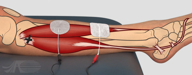
Electrode configuration for ankle dorsiflexion. The fibular head is marked, and the lateral malleolus is visible. An asymmetric biphasic waveform is used, with the negative electrode placed over the muscle belly of the anterior tibialis, very midline close to the tibia.
The positive electrode is also placed close to the tibia, further down the shank.
Related Electrode Placements
Ankle Dorsiflexion
Video Instruction
Audio Transcript:
Electrode configuration for ankle dorsiflexion. The fibular head is marked, and the lateral malleolus is visible. An asymmetric biphasic waveform is used, with the negative electrode placed over the muscle belly of the anterior tibialis, very midline close to the tibia.
The positive electrode is also placed close to the tibia, further down the shank. During stimulation a three out of five contraction can be seen, with good balance of the foot and minimal activation of the toe extensors.
Ankle Dorsiflexion
Muscle Anatomy

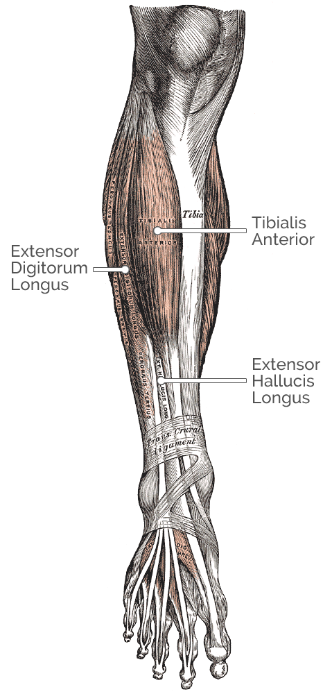
Muscles involved in ankle dorsiflexion:
Tibialis Anterior
Origin: Upper 1/2 or 2/3 of the lateral surface of the tibia and the adjacent interosseous membrane.
Insertion: Medial cuneiform and the base of first metatarsal bone of the foot
Other actions: Foot inversion
Extensor Hallucis Longus
Origin: Arises from the middle portion of the fibula on the anterior surface and the interosseous membrane.
Insertion: Inserts on the dorsal side of the base of the distal phalanx of the big toe.
Other actions: Extends big toe
NOTE: The activation of this muscle with the TA is not advantageous, as active toe extension within a shoe can lead to discomfort and skin breakdown
Extensor Digitorum Longus
Origin: Arises from the middle portion of the fibula on the anterior surface and the interosseous membrane.
Insertion: Inserts on the dorsal side of the base of the distal phalanx of the big toe.
Other actions: Extension of toes.
NOTE: The activation of this muscle with the TA is not advantageous, as active toe extension within a shoe can lead to discomfort and skin breakdown
Ankle Dorsiflexion
Nerve Anatomy

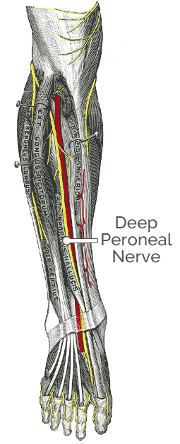
Nerves involved in ankle dorsiflexion:
Tibialis Anterior
Nerve innervation: Peroneal nerve Nerve root: L5
Extensor Hallucis Longus
Nerve innervation: Deep peroneal nerve, branch of common peroneal nerve
Nerve root: L4, L5, S1
Extensor Digitorum Longus
Nerve innervation: Deep peroneal nerve
Nerve root: L5
This website uses cookies to analyze site traffic and provide you with the best experience possible. Learn more
This website uses cookies to analyze site traffic and provide you with the best experience possible.
Learn more
